Powering Digital Pathology 2.0
Pramana’s Technology
Intelligent scanners. Enriched Images. Confidence for pathologists, lab directors, and technicians. Learn about what makes our technology stand out!
Pramana Partner Program - Validated IMS Partners
Pramana is leading the industry transformation with Digital Pathology 2.0. We are excited about our high value partnerships standardizing WSI data through DICOM.

PathPresenter is the image sharing platform for pathology, on a mission to democratize access to the world’s pathology knowledge by connecting pathologists to the vast expertise of their colleagues. PathPresenter Exchange is designed to streamline the process of sharing digital pathology images and data across multiple locations and users. The software includes features such as image annotation, case collaboration, and reporting tools to facilitate communication and collaboration among users.

Gestalt transforms pathology through an intelligent, configurable, and vendor-neutral digital workflow that provides true interoperability to enable pathologists. Gestalt’s PathFlow Viewer is designed to provide pathologists and researchers with a comprehensive digital pathology solution that includes image management, analysis, and collaboration tools.
Corista delivers the industry’s most extensive array of workflow, analytical and collaborative tools for pathology. Their platform enables the storage, viewing, analysis, and sharing of digital pathology images and data, allowing for faster and more efficient diagnoses and research.
Autonomous Operations and Error Elimination
Our solutions address variability across the entire digital pathology workflow. With most scanners on the market, manual processing of slides such as focal point selection, QA/QC and re-running problem slides is still required, resulting in variability. Labs are forced to employ skilled technicians throughout the digital operation to overcome the technology limitations.
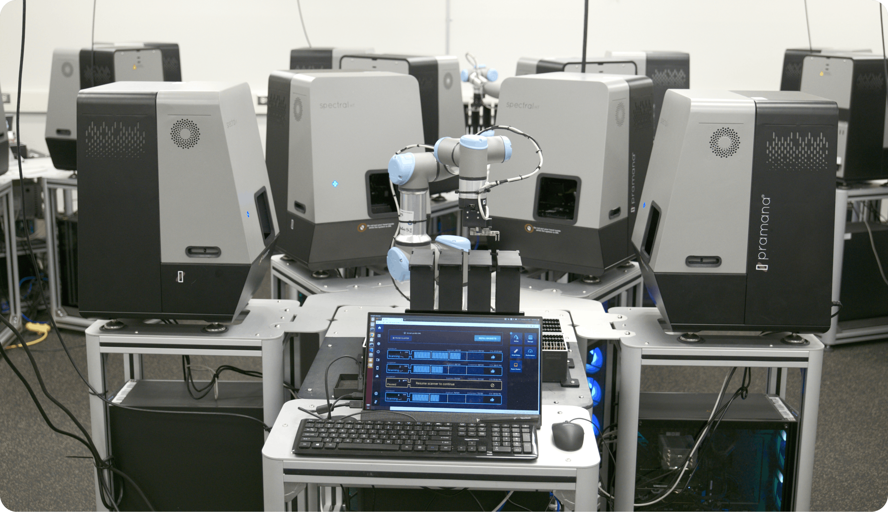
Digital Pathology 2.0 presents a major step forward in whole slide imaging. Pramana’s software and algorithms automate scan parameter setting and real-time error detection and correction, eliminating variability and increasing confidence through superior quality WSIs. Furthermore, pathologists don’t always concur with each other while reading glass slides under a microscope. It has been proven that this discordance gets amplified, or at best, remains the same when interpreting digital images. However, Digital Pathology 2.0 brings the opportunity to change this reality. By enriching the images and applying novel visualization tools, the inter-observer pathologist-level variability to assess a case is being addressed.
Common Artifacts are Handled by Pramana's Pixel Level Z-stack Focusing Technology
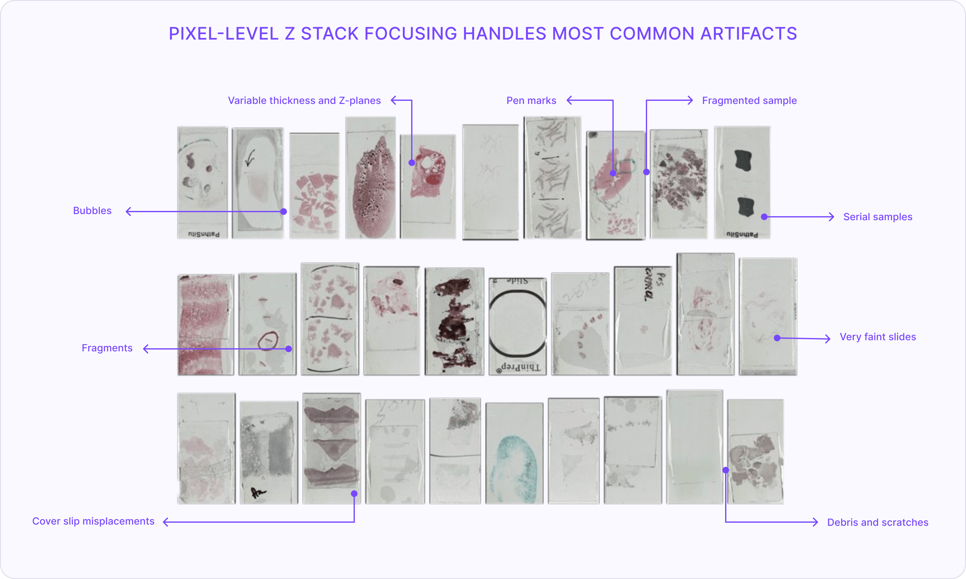
Our scanners perform extremely fast focus sampling and dynamically calculate the z plane of the tissue. The scanners make in-line decisions based on tissue type, thickness and slide artifacts like annotations and debris.
Whole Slide Image (WSI) Analytics
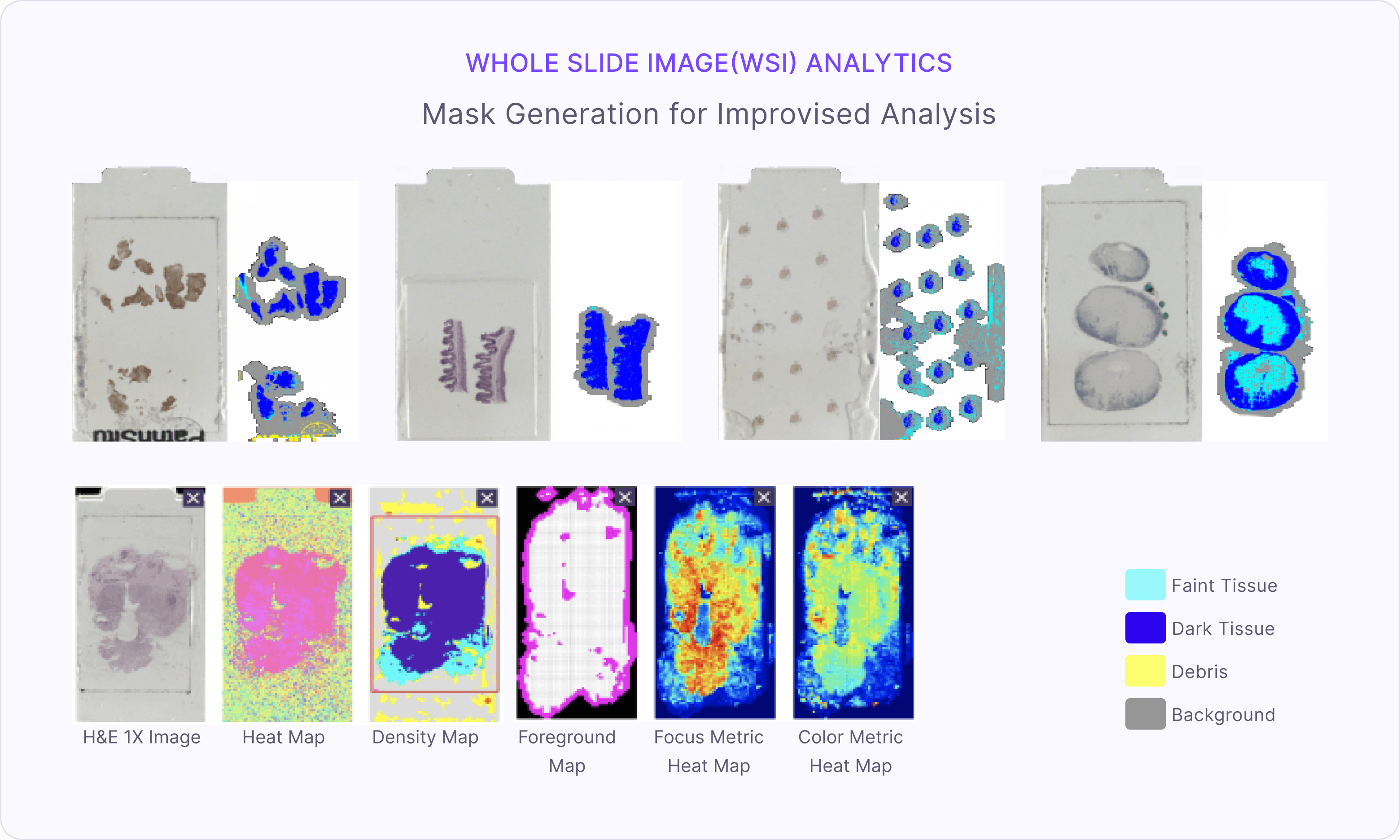
While the acquired images are processed for accurate focus, they are also embedded with a wealth of image quality and analytics data. Select features, including focus and stitching, are abstracted to tile-based feature overlays. The feature overlays provide an “at a glance” snapshot of slide quality and can also be integrated into a numeric quality metric (if desired) to drive lab and technician performance or downstream quality control activities.
Standardized Image and Data Formats with DICOM Support
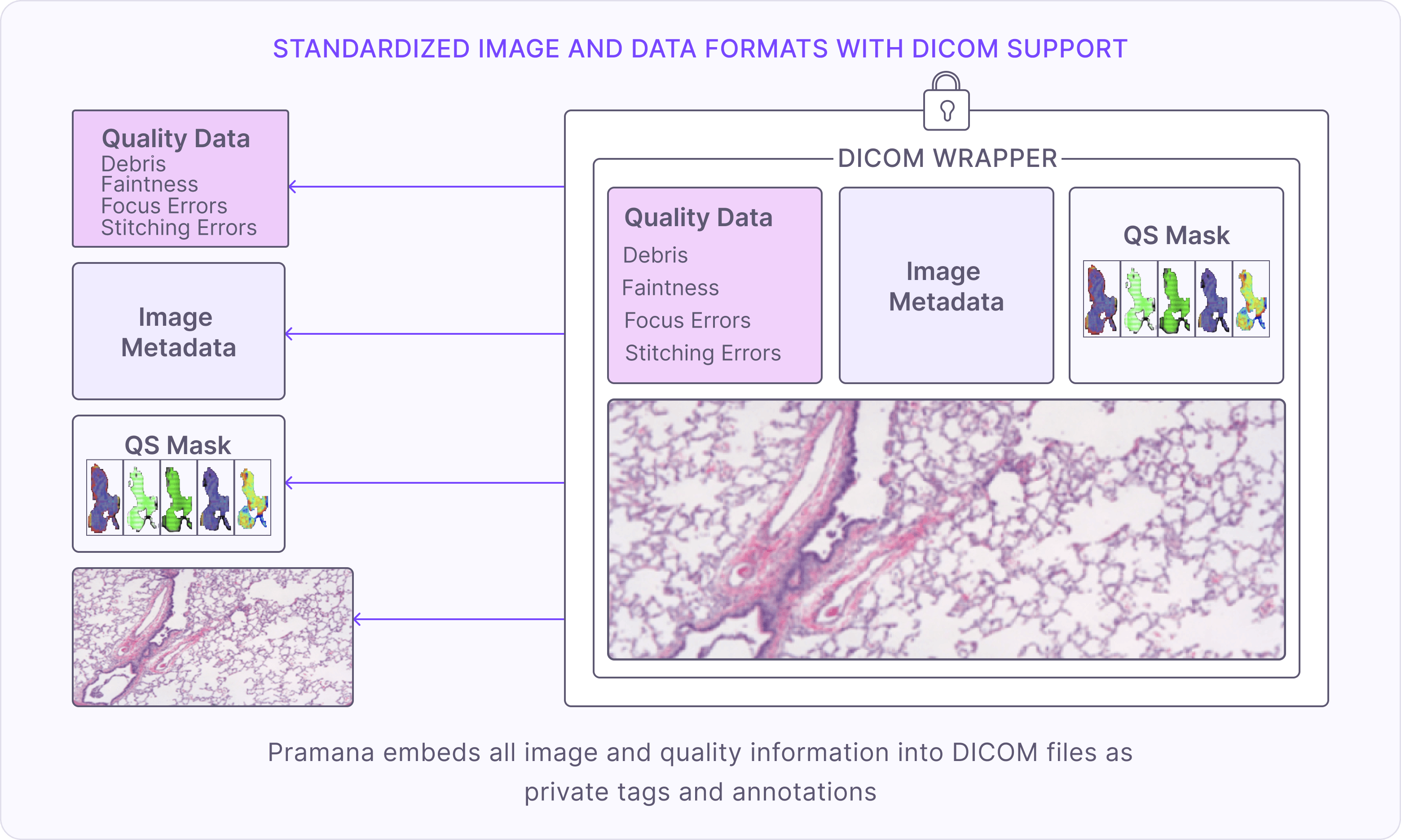
Our scanners output data in industry standard formats like DICOM (Composite, multi-level) and OME-TIFF (composite, multi-level) that allow for interoperability across various external system and image viewers. The whole slide images and corresponding metadata have been successfully submitted to interoperability tests run by industry bodies and have been integrated with some of the leading image management utilities for viewing in 3rd party viewers.