Check out our Posters from European Congress of Pathology 2024

BrochureBrochure

Autonomous

Whole Slide Imaging Systems

Digital Pathology 2.0 from Pramana
Economical
Precise
Stress-free
mayoDukeCarisironMarupIMmayoDukeCarisironMarupIM
Partnering With Leading Healthcare Organizations
img

The need for a new approach

icon
Inherent Challenges with Slides

Many scanners fail to effectively manage slide loading, label handling, focus point selection, and artifact handling, leading to an increased need for operator adjustments.

icon
Manual Quality Control

High error rates for daily specimens place a significant burden on trained operators, requiring them to manually identify errors and determine appropriate next steps.

icon
Under-Utilized Scanner Capacity

Most labs load only 60-80 slides per batch, yet they are required to invest in capacity of 300-1000 slides. Despite this, just one slide is scanned at a time leading to a significant underutilization.

icon
Cloud Image Analysis Challenges

Cloud-based image analysis is restrictive, costly, and slow, requiring gigabyte transfers for even simple algorithms and typically processing images 3-4 times slower than the actual scanning time.

img
img

Spectral family of scanners
from Pramana

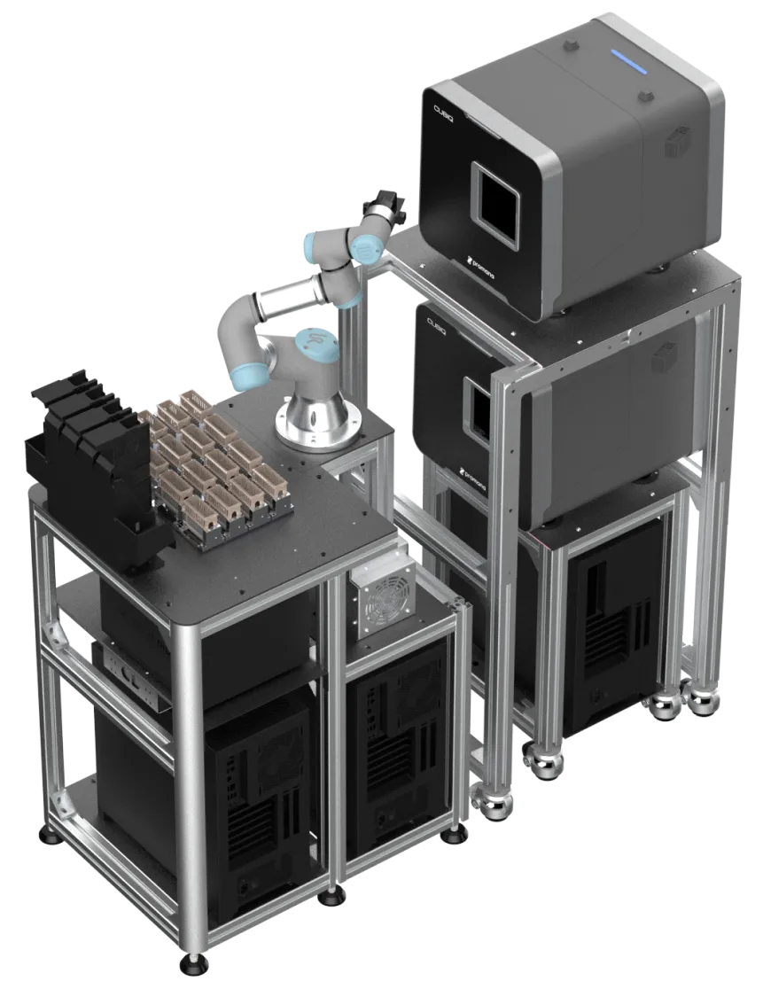
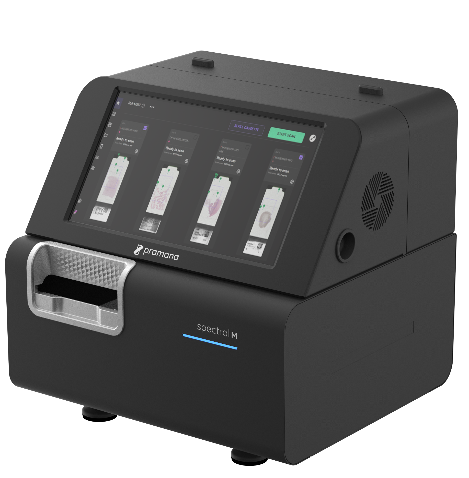
Pramana's modular scanner systems feature identical optical paths, uniform software, and a seamless user experience.

As needs grow, additional scanner heads can be integrated effortlessly. By separating the slide handling mechanism from the scan heads, Pramana offers labs an innovative approach to design tailored solutions that meet their capacity requirements.
Pramana's modular scanner systems feature identical optical paths, uniform software, and a seamless user experience.
As needs grow, additional scanner heads can be integrated effortlessly.By separating the slide handling mechanism from the scan heads, Pramana offers labs an innovative approach to design tailored solutions that meet their capacity requirements.
img
img

A generational advancement in

Digital Pathology from Pramana

solution icon
Autonomous Operation
Handle most slide types without any user intervention, including challenging slides
solution icon
Modular and Scalable Solutions
Modular systems for quick capacity upgrades
Pramana's solutions
solution icon
Built-in QA
Inline quality assurance with fully autonomous rescanning(wherever needed)
solution icon
Edge AI
Capability to execute multiple feature description algorithms in real time during the scanning process.
Pramana's solutionsPramana's
Solutions
solution icon
Autonomous Operation
Handle most slide types without any user intervention, including challenging slides
solution icon
Built-in QA
Inline quality assurance with fully autonomous rescanning(wherever needed)
solution icon
Modular and Scalable Solutions
Modular systems for quick capacity upgrades
solution icon
Edge AI
Capability to execute multiple feature description algorithms in real time during the scanning process.
img
img

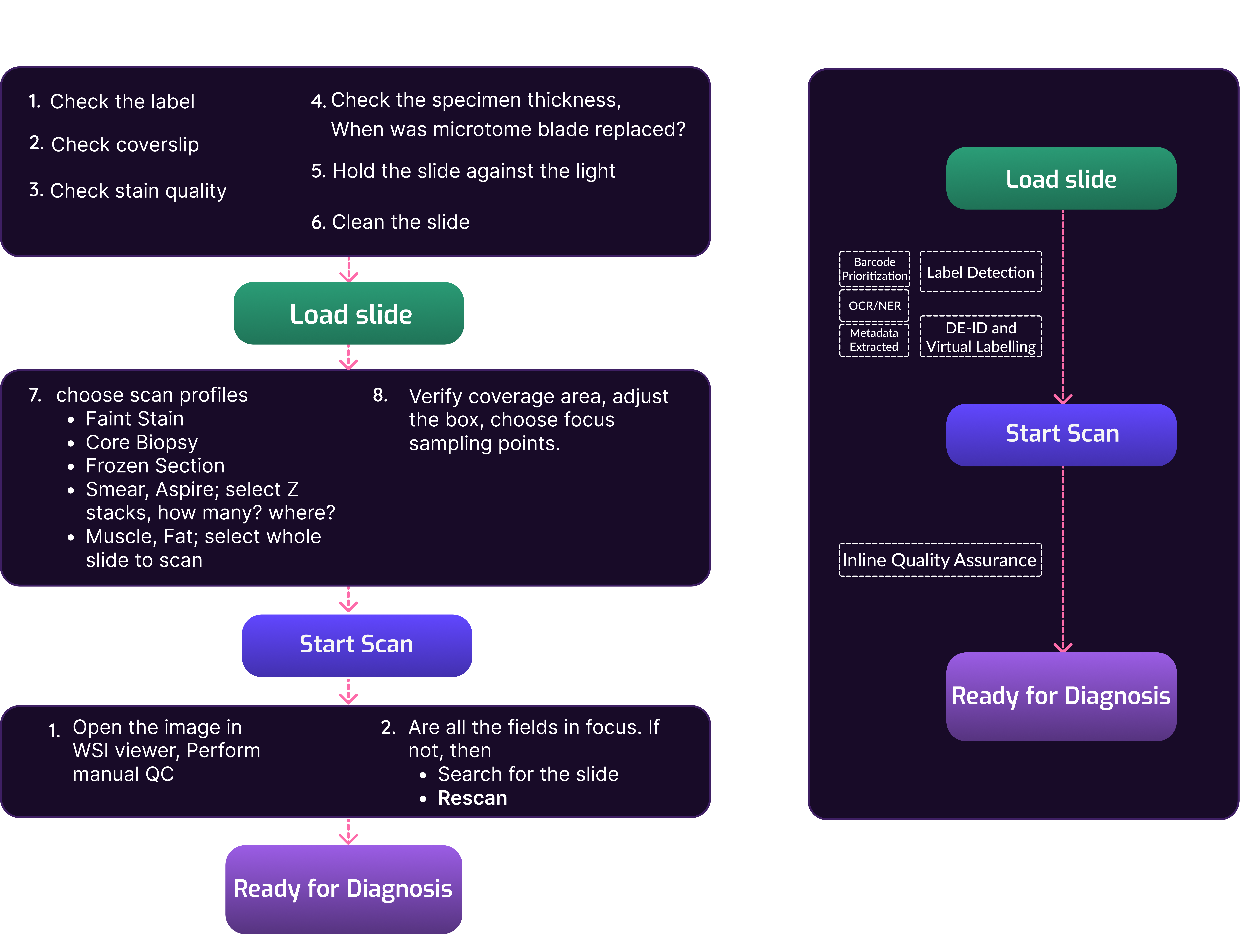
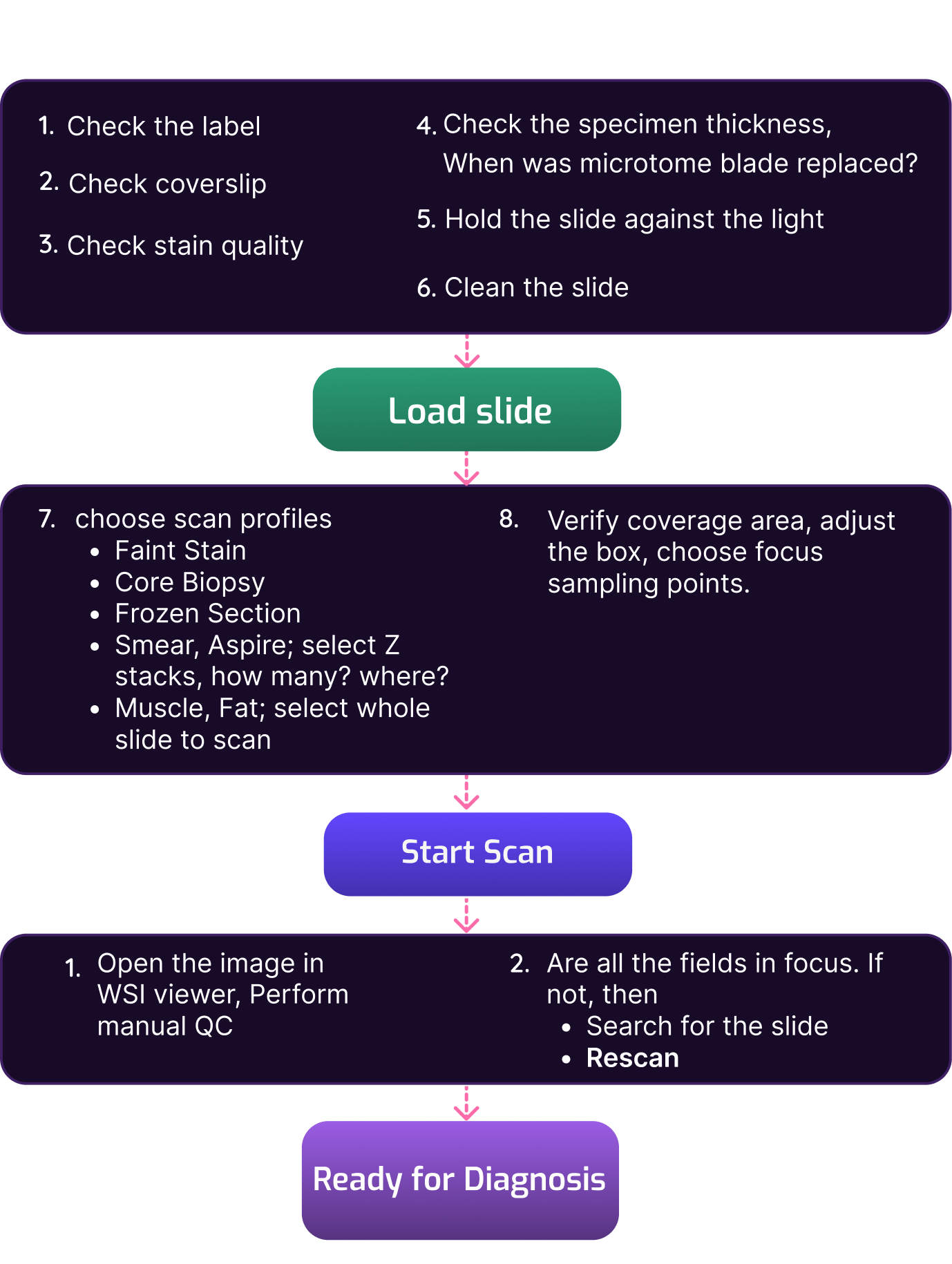
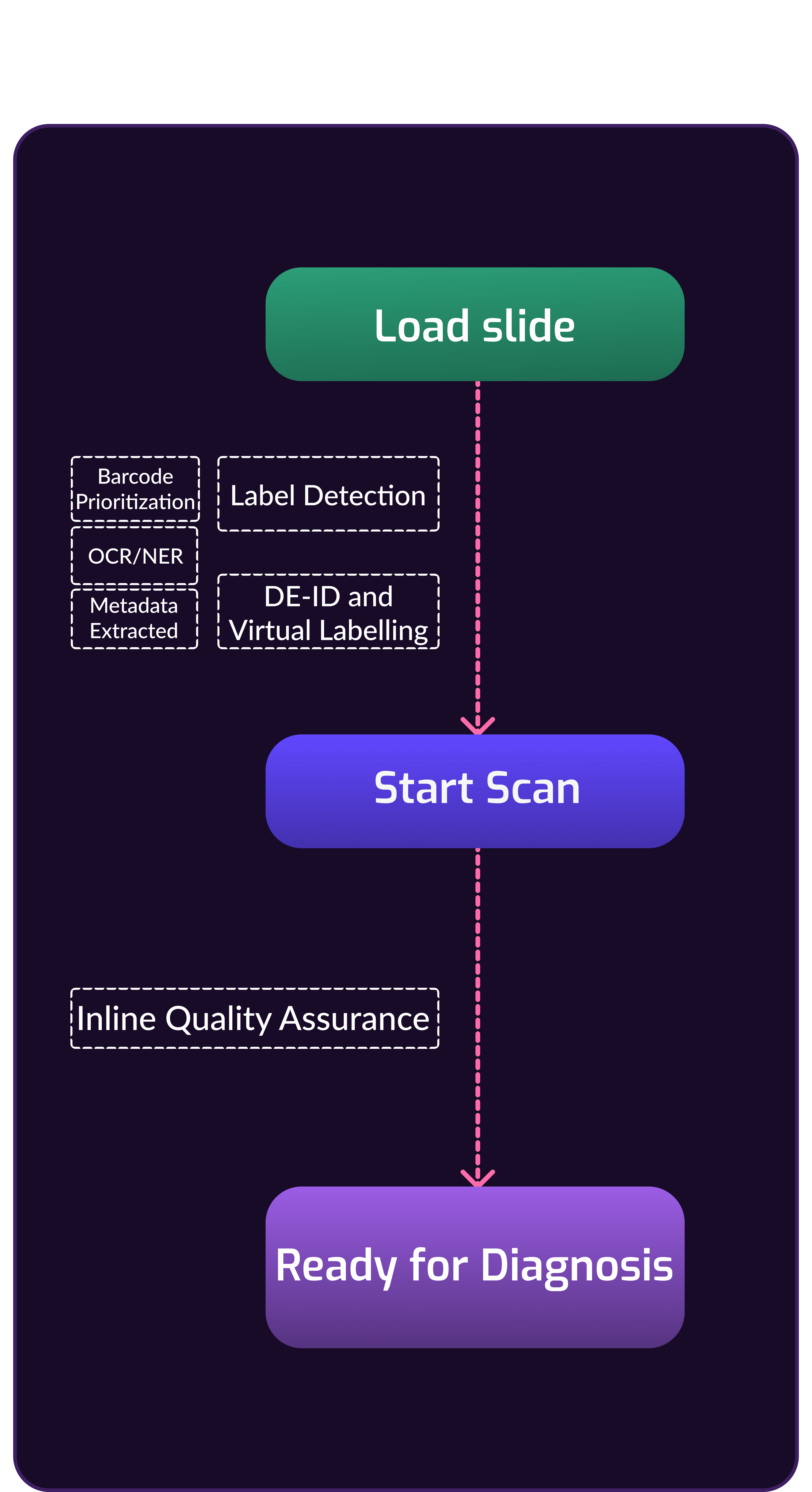
Autonomous imaging looks like this

process flow diagram to show difference between other scanners and pramana scannersprocess flow diagram of other scannersprocess flow diagram of pramana scanners

Product Configurations

Each lab has its own approach to optimizing layout and workflow, with varying upstream systems such as stainers, coverslip techniques, and drying racks. Pramana allows customers to begin with a base design and customize their setup by selecting the layout, number of scan heads, and slide baskets according to their specific needs. Additionally, custom kits are available to accommodate future changes as desired.
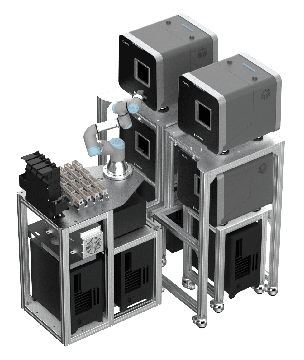
SpectralHT 4-Head
SpectralHT 4-Head
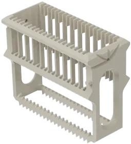
Basket image
960 Slides
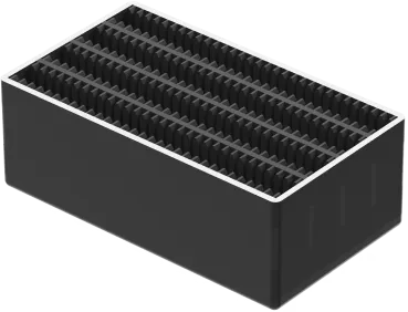
8 Pramana Baskets
480 Slides
4 Pramana Baskets
Basket image
320 Slides
16 sakura Baskets

SpectralHT 2-Head
SpectralHT 2-Head
Basket image
480 Slides
4 Pramana Baskets
240 Slides
2 Pramana Baskets
Basket image
320 Slides
16 sakura Baskets

SpectralM
SpectralM
Basket image
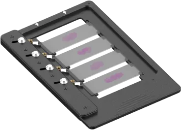
4 Slides
1''x3'' slides
Basket image
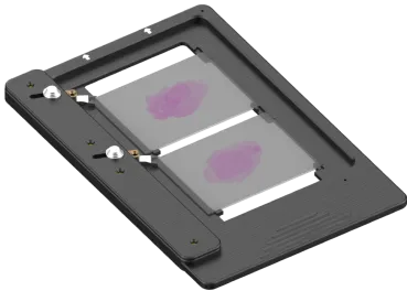
2 Slides
2''x3'' slides
img

Specifications

ApplicationsAnatomic Pathology, Cytology, Microbiology, Archives, Frozen Sections
Specimen TypeTissue, Frozen Sections, Bone Marrow, Smears
Average Scan Time Per Load6-7 hours for 480 slide load
WSI Output FormatDICOM, OME - TIFF, DZI
Magnifications4x, 20x, 40x, 80x
Resolution0.25 microns per pixel at 40x
Slide Artifact HandlingVery faint tissue, very tiny tissue, thick cuts, double thick slides, broken coverslips, debris, dirty coverslips, protruding labels
Image compressionJPEG Visually Lossless (configurable), ZStandard
img

Specimen Compatibility

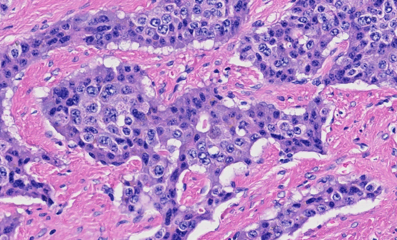
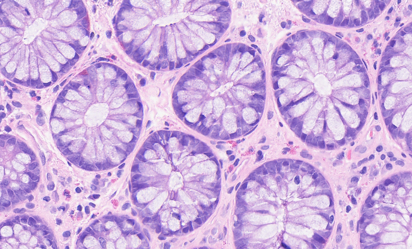
If a glass slide is approved by the pathologist, Pramana’s system will likely generate a high-quality whole slide image without any errors, regardless of the specimen type, including tissue, cytology, bone marrow, microbiology, and wet mount samples.
Tissue
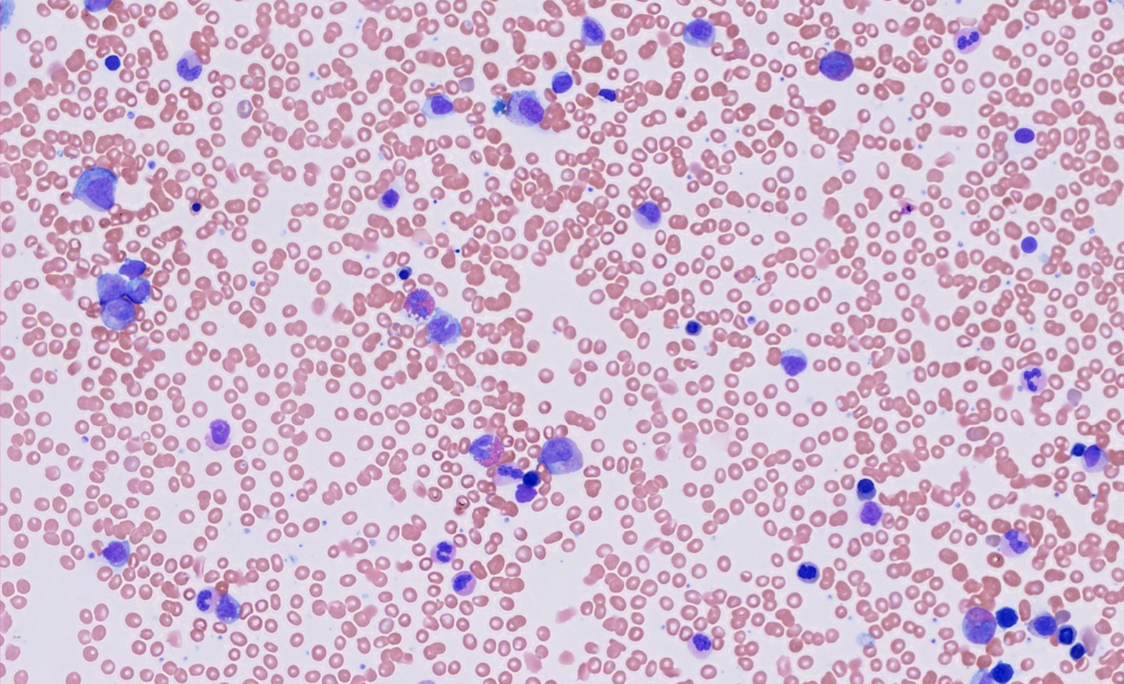
Cytology
Bone Marrow
Microbiology
Wet Mount
IDC breast Image
IDC breastH&E, 40X
Intestine Image
IntestineH&E, 40X
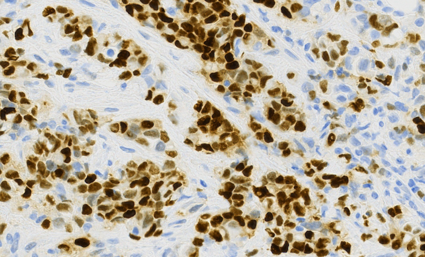
IHC Image
IHCER, 40X
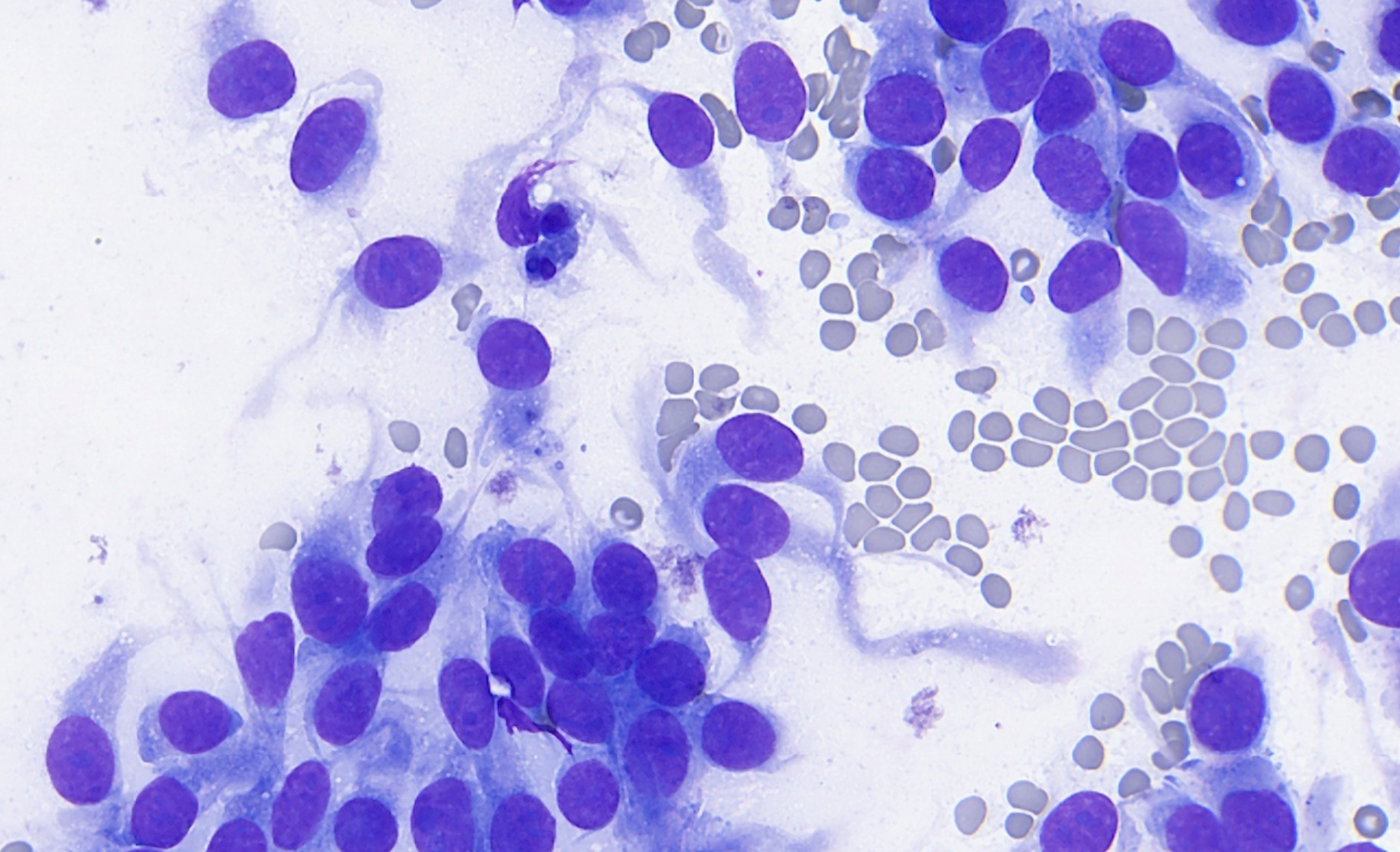
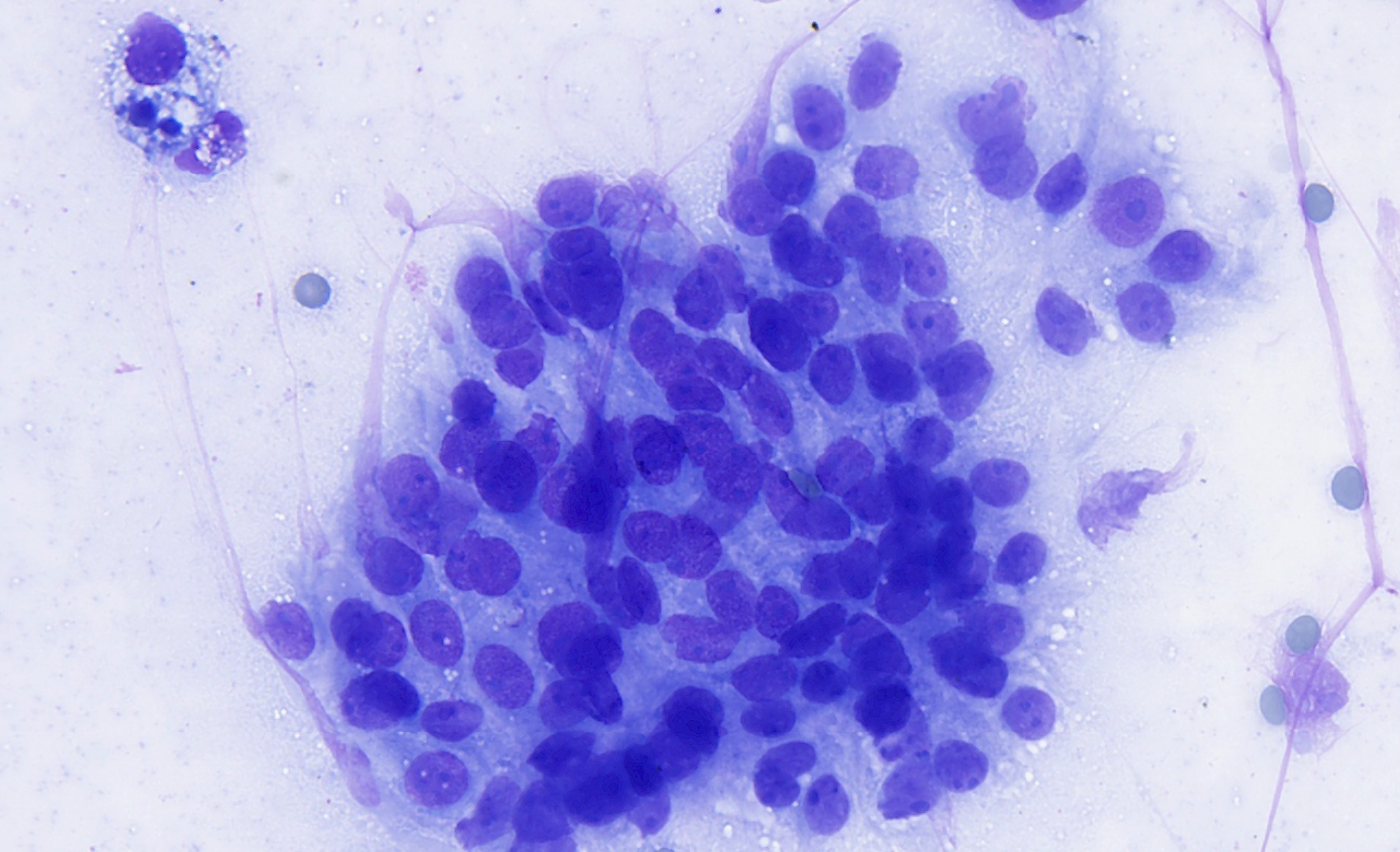
Cytology conventional smear Image
Cytology conventional smearMGG stain, 40X
Cytology conventional smear Image
Cytology conventional smearMGG stain, 40X
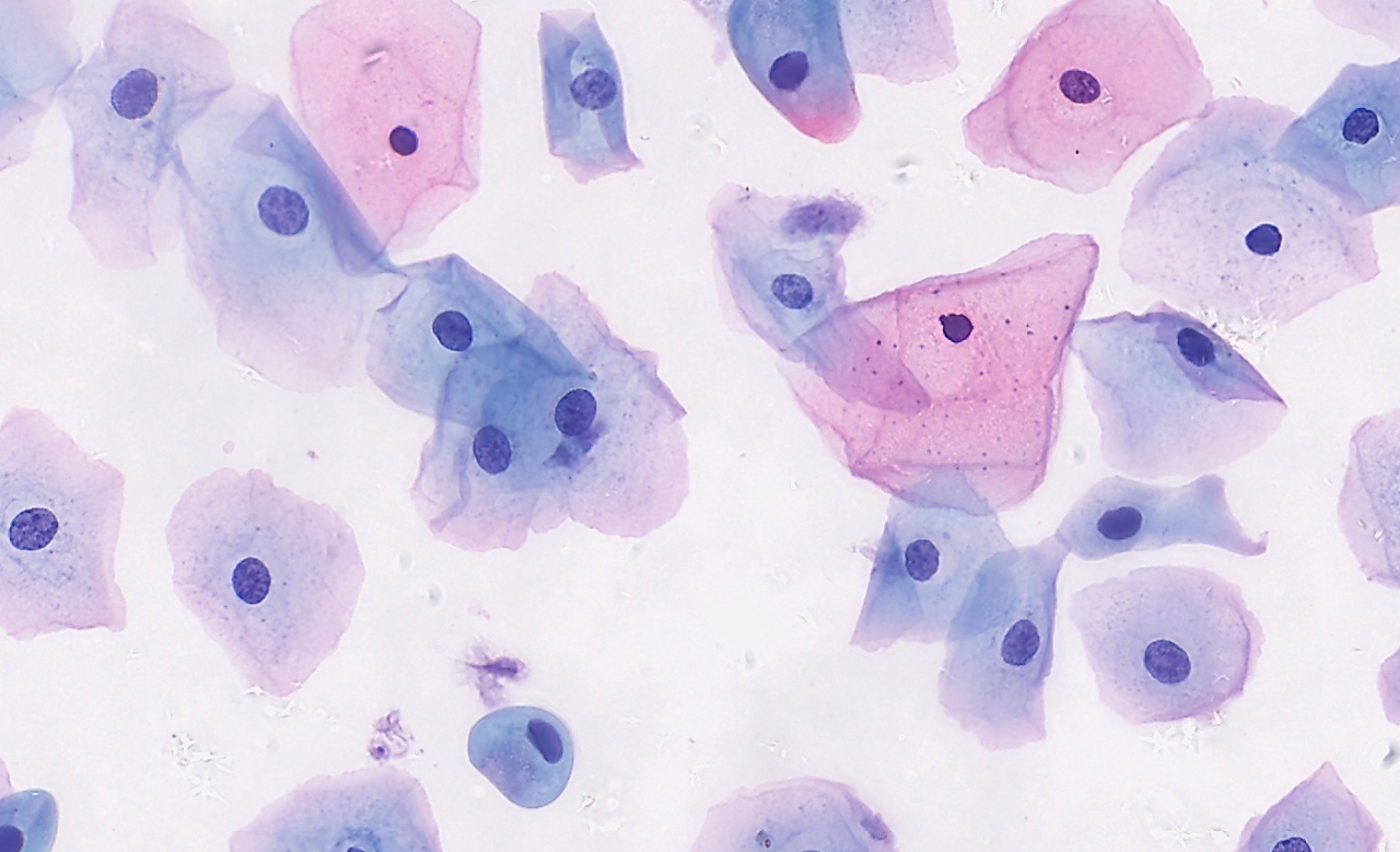
Liquid based Cytology (LBC) Image
Liquid based Cytology (LBC)PAP stain, 40X
Bone marrow aspirate smear Image
Bone marrow aspirate smearGiemsa stain, 40X
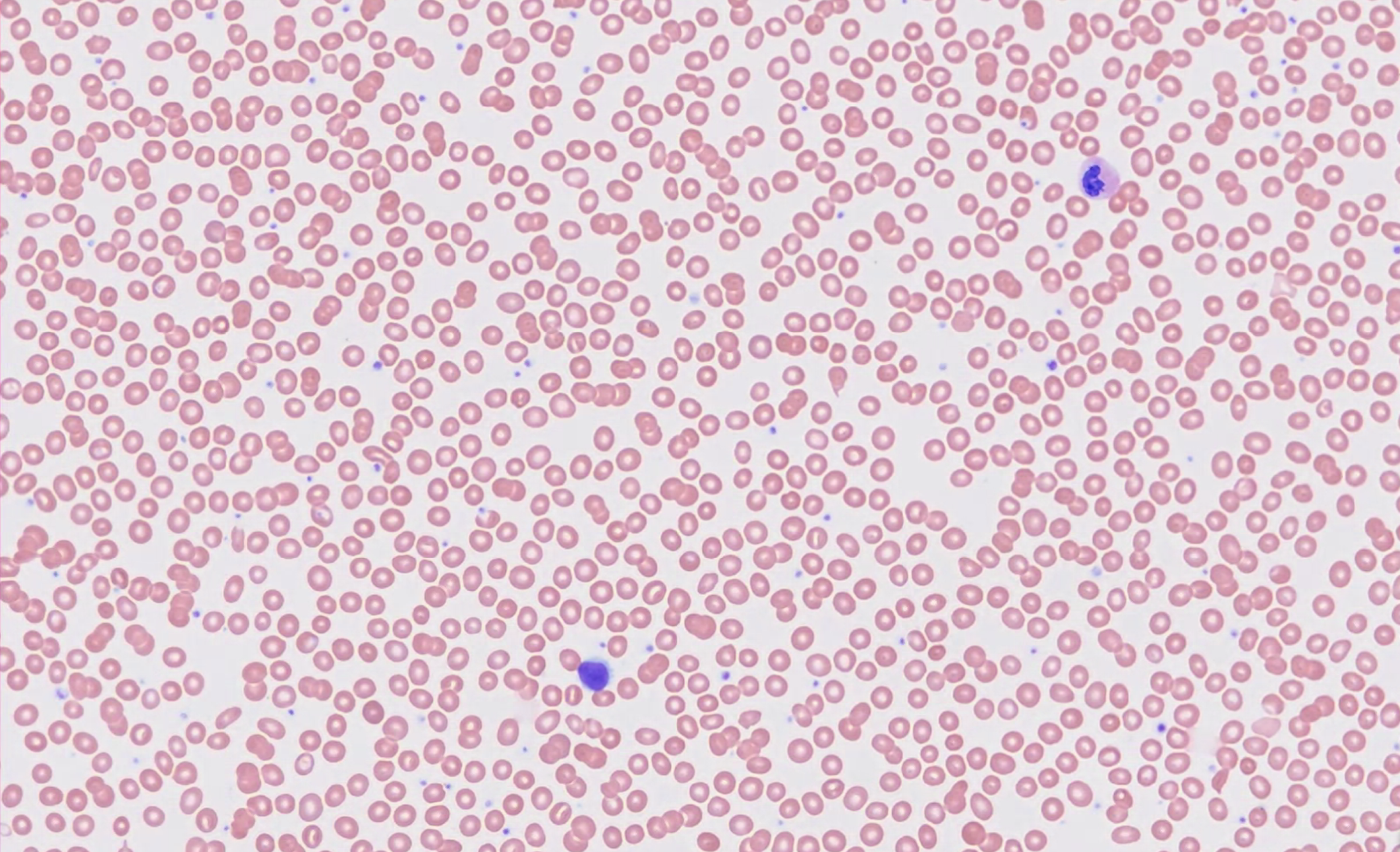
Peripheral smear Image
Peripheral smearGiemsa stain, 40X
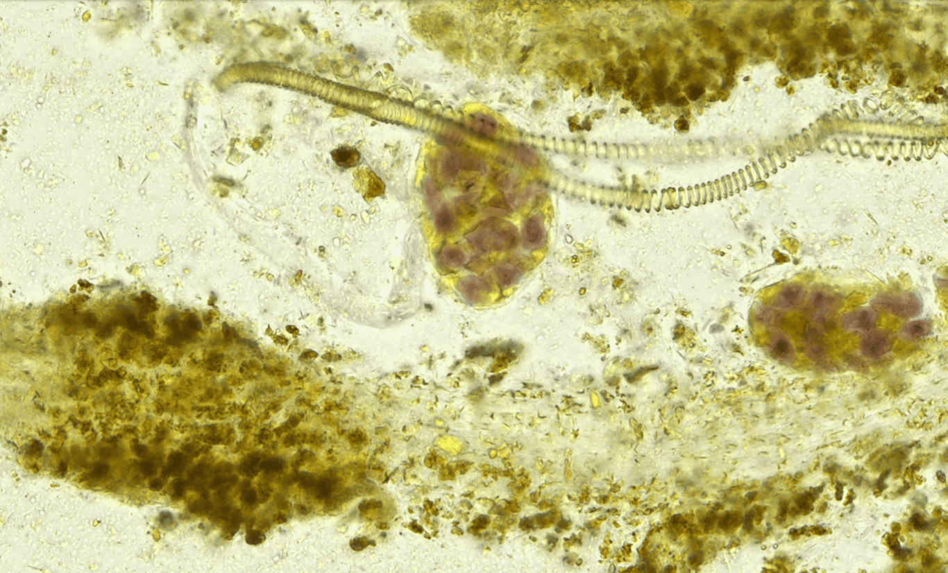
Human fecal trichrome Image
Human fecal trichrome40X
img

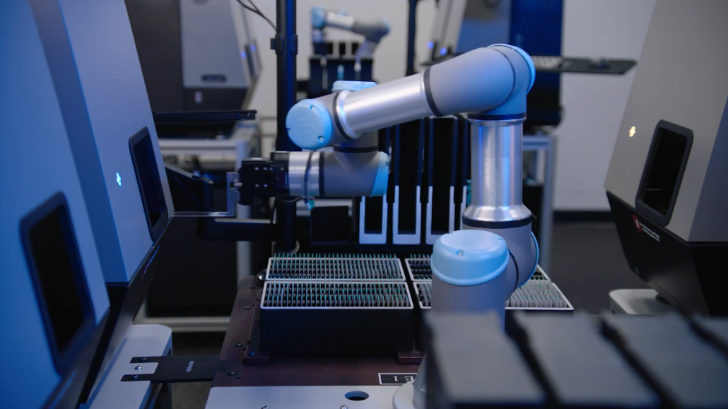
Our scanners in action

Watch Pramana Scanners in<br/> actionPlay button
Watch Pramana Scanners in
action
img

How do we do it ?

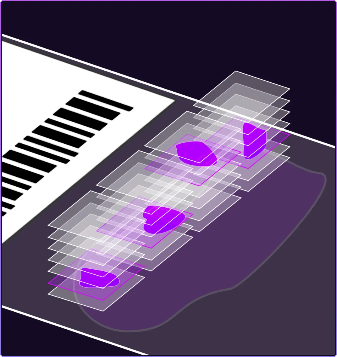
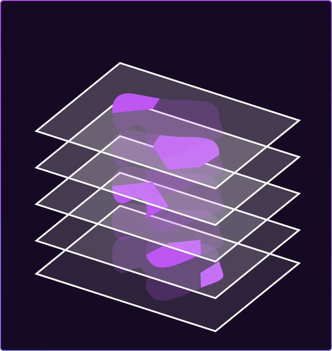
By replicating the fine-focus function of an optical microscope, Pramana has pioneered a groundbreaking method to volumetric imaging that enhances image quality and detail while improving scanning efficiency.

Backed by a rich portfolio of granted U.S. patents

img

Volumetric Imaging

Volumetric Imaging process image

Volumetric scanning at each
field of view

next step
Volumetric Imaging process image

Fusion of Z-stacks and stitching

next step
Volumetric Imaging process image

Fully focused image

img
img

Inline Computing leading to Edge AI

Real-time algorithms for feature detection, quantification, and classification enable inline computation during scanning, ensuring that results are instantly available upon the scan's completion.

This approach optimizes efficiency, reduces costs, and enhances safety during the scanning process. Additionally, the ability to securely integrate third-party algorithms within the scanner significantly elevates the value of digital pathology for both research and clinical use.

img
Proscia iconGSTALT iconpathPresenter icontechcyte iconcorista icon
Interoperable with IMS Vendors

Conformité Européenne certification iconThe European In Vitro Diagnostic Devices Regulation certificationiso certification
In U.S., Available for Research Use Only

News and Announcements

poster of the news or announcements
Spectrum Healthcare Partners Leverages Pramana’s AI-Enabled Scanners to Bring Digital Pathology to Central Maine Healthcare
2024-10-24
READ MORE
click to visit the page
poster of the news or announcements
Pramana Achieves Industry-Leading Milestone with Digital Pathology 2.0 Technology
2024-03-22
READ MORE
click to visit the page
poster of the news or announcements
Pramana Joins Proscia Ready To Help Laboratories Realize More Value From Their Pathology Data
2024-02-27
READ MORE
click to visit the page

Join the Digital Pathology 2.0 Revolution

Pramana is transforming digital pathology through its.
hardware-software co-design for whole slide imaging.